Anatomy and Physiology
To understand bacterial meningitis, we should first understand the related anatomy and physiology of a healthy individual.
Meningitis, in general, is the inflammation of the protective membranes surrounding the brain and spinal cord. In the case of bacterial meningitis, this inflammation is caused by bacterial infection. In order to inflame these protective membranes, the bacteria must somehow enter the bloodstream and bypass the blood-brain barrier.
Meningitis, in general, is the inflammation of the protective membranes surrounding the brain and spinal cord. In the case of bacterial meningitis, this inflammation is caused by bacterial infection. In order to inflame these protective membranes, the bacteria must somehow enter the bloodstream and bypass the blood-brain barrier.
Blood-Brain Barrier
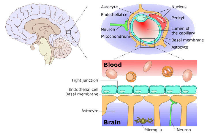
Since the brain is such a delicate organ, nature has taken extra measures to protect the brain by creating the blood-brain barrier to limit the diffusion of substances from the bloodstream into brain tissue selectively.
The blood-brain barrier mainly consists of tight junctions, which seals the endothelial cells that line the brain capillaries. Astrocytes, a type of neuroglia from the brain, closely attached to the endothelial cells and release chemicals to regulate the permeabilities of the tight junctions. The major sites of the blood brain barrier are the arachnoid membrane, choroid plexus epithelium, and the cerebral microvascular endothelium.
Only a few kinds water-soluble substance can move across the blood-brain barrier, such as glucose by active transport, urea, creatinine, and ions move across by slow diffusion. On the other hand, lipid-soluble substances can easily cross the blood-brain barrier, such as oxygen, carbon dioxide, alcohol, and most anesthetic agents.
When bacteria break through the blood-brain barrier, an infection occurs in the cerebrospinal fluid.
Blood-Brain Barrier [1]
Cerebrospinal Fluid
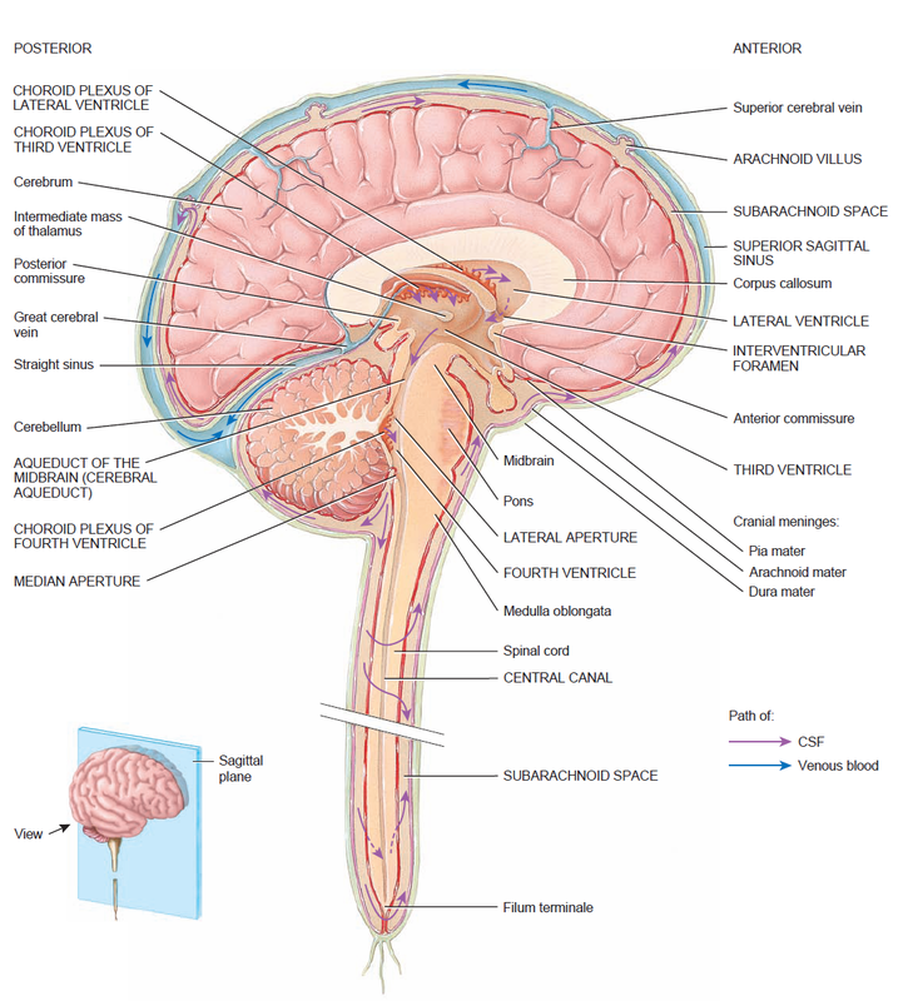
Cerebrospinal fluid (CSF) is a colorless, transparent liquid that continuously circulates through the cavities of the brain and spinal cord, and as such, it acts as an internal circulation system to transport nutrients and wastes between the bloodstream and the brain and spinal cord. This reducdant circulation protects the brain and spinal cord from chemical injuries similar to the function of the blood-brain barrier. The CSF also protects the brain and spinal cord from physical injuries by acting as a shock absorber between the brain and spinal cord from the skeletal structures (cranium and vertebrae) [2].
CSF is produced in the choroid plexuses, which are networks of capillaries in the ventricles. The choroid plexuses filter out blood plasma from the bloodstream, which is the main component of CSF. The choroid plexuses are covered by ependymal cells that are sealed together with tight junctions. These tight junctions forces the blood plasma to pass through these ependymal cells, which further filter out the blood plasma, producing CSF [2].
From the choroid plexuses of each lateral ventricle, CSF flows into the third ventricle through the interventricular foramina, which are two narrow oval openings. The choroid plexuses in the third ventricle adds more CSF. Then, CSF flows into the fourth ventricle throught the cerebral aqueduct. Again, the choroid plexuses in the fourth ventricle adds more CSF. The fluid then enters the subarachnoid spacethrough the three openings in the roof of the fourth ventricle. These three openings are a median aperture and a pair of lateral apertures. Then, CSF circultates in the central canal of the spinal cord and in the subarachnoid space around the surface of the brain and spinal cord [2].
CSF Circulation [2]
Meninges
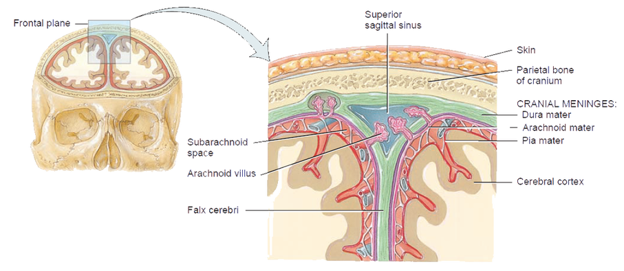
The meninges are three connective tissue coverings that encircle the spinal cord and brain. The spinal meninges surround the spinal cord and are continuous with the cranial meninges, which encircle the brain [2].
The meninges lined the cranial and vertebral cavities to protect the brain and the spinal cord, and they are also attached to the cranial bones' inner surfaces, which facilitate the crainal bones to stabilize the positions of the brain, blood vessels, lymphatic vessels, and nerves [2].
These three connective tissue coverings are dura mater, which is composed of dense, irregular connective tissue), arachnoid mater, which is composed of delicate collagen fibers and some elastic fibers in a spider’s web arrangement, and pia mater, which is a thin transparent connective tissue layer consists of squamous to cuboidal cells within interlacing bundles of collagen fibers and some fine elastic fibers [2].
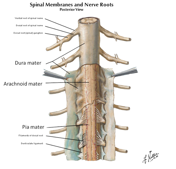
The most superficial of the three spinal meninges, the dura mater, forms a sac from the level of the foramen magnum in the occipital bone, where it is continuous with the dura mater of the brain, to the second sacral vertebra. The spinal cord is also protected by a cushion of fat and connective tissue located in the epidural space, a space between the dura mater and the wall of the vertebral canal [2].
The middle meninx is an avascular covering called the arachnoid mater. It is deep to the dura mater and is continuous with the arachnoid mater of the brain. Between the dura mater and the arachnoid mater is a thin subdural space, which contains interstitial fluid [2].
The innermost meninx is the pia mater, which adheres to the surface of the spinal cord and brain. Within the pia mater are many blood vessels that supply oxygen and nutrients to the spinal cord. Between the arachnoid mater and the pia mater is the subarachnoid space, which contains cerebrospinal fluid that serves as a shock absorber and suspension system for the spinal cord and brain [2].
All three spinal meninges cover the spinal nerve roots, structures that connect spinal nerves to the spinal cord, up to the point where they exit the spinal column through the intervertebral foramina. Triangular-shaped membranous extensions of the pia mater suspend the spinal cord in the middle of its dural sheath. These extensions, called denticulate ligaments, are thickenings of the pia mater. They project laterally and fuse with the arachnoid mater and inner surface of the dura mater between the anterior and posterior nerve roots of spinal nerves on either side. Extending all along the length of the spinal cord, the denticulate ligaments protect the spinal cord against sudden displacement that could result in shock [2].
The meninges lined the cranial and vertebral cavities to protect the brain and the spinal cord, and they are also attached to the cranial bones' inner surfaces, which facilitate the crainal bones to stabilize the positions of the brain, blood vessels, lymphatic vessels, and nerves [2].
These three connective tissue coverings are dura mater, which is composed of dense, irregular connective tissue), arachnoid mater, which is composed of delicate collagen fibers and some elastic fibers in a spider’s web arrangement, and pia mater, which is a thin transparent connective tissue layer consists of squamous to cuboidal cells within interlacing bundles of collagen fibers and some fine elastic fibers [2].
The most superficial of the three spinal meninges, the dura mater, forms a sac from the level of the foramen magnum in the occipital bone, where it is continuous with the dura mater of the brain, to the second sacral vertebra. The spinal cord is also protected by a cushion of fat and connective tissue located in the epidural space, a space between the dura mater and the wall of the vertebral canal [2].
The middle meninx is an avascular covering called the arachnoid mater. It is deep to the dura mater and is continuous with the arachnoid mater of the brain. Between the dura mater and the arachnoid mater is a thin subdural space, which contains interstitial fluid [2].
The innermost meninx is the pia mater, which adheres to the surface of the spinal cord and brain. Within the pia mater are many blood vessels that supply oxygen and nutrients to the spinal cord. Between the arachnoid mater and the pia mater is the subarachnoid space, which contains cerebrospinal fluid that serves as a shock absorber and suspension system for the spinal cord and brain [2].
All three spinal meninges cover the spinal nerve roots, structures that connect spinal nerves to the spinal cord, up to the point where they exit the spinal column through the intervertebral foramina. Triangular-shaped membranous extensions of the pia mater suspend the spinal cord in the middle of its dural sheath. These extensions, called denticulate ligaments, are thickenings of the pia mater. They project laterally and fuse with the arachnoid mater and inner surface of the dura mater between the anterior and posterior nerve roots of spinal nerves on either side. Extending all along the length of the spinal cord, the denticulate ligaments protect the spinal cord against sudden displacement that could result in shock [2].
Cranial Meninges [2]
Spinal Cord Meninges [3]
References
[1] Netter, F. H. (2006). Atlas of Human Anatomy (4th ed.). Philadelphia: Saunders Elsevier.
[2] Simkó, M., Fiedeler, U., Gazsó, A., & Nentwich, M. (2010, December). Can nanoparticles end up in the brain? NanoTrust Dossiers(014en).
[3] Tortora, G. J., & Derrickson, B. (2009). Principles of Anatomy and Physiology (12th ed.). Danver, United States of America: John Wiley & Sons, Inc.
[2] Simkó, M., Fiedeler, U., Gazsó, A., & Nentwich, M. (2010, December). Can nanoparticles end up in the brain? NanoTrust Dossiers(014en).
[3] Tortora, G. J., & Derrickson, B. (2009). Principles of Anatomy and Physiology (12th ed.). Danver, United States of America: John Wiley & Sons, Inc.
